CLL chromosome and gene anomaly detection probe (P53/CEP17)
P53 Gene Probe Detection Kit - 100µL/10 Tests
TP53 mutations are universal across cancer types. The loss of a tumor suppressor is most often through large deleterious events, such as frameshift mutations, or premature stop codons. In TP53 however, many of the observed mutations in cancer are found to be single nucleotide missense variants. These variants are broadly distributed throughout the gene, but with the majority localizing in the DNA binding domain. There is no single hotspot in the DNA binding domain, but a majority of mutations occur in amino acid positions 175, 245, 248, 273, and 282 (Olivier et al., 2010).
To fulfill its proper biological function four TP53 polypeptides must form a tetramer which functions as a transcription factor, therefore even if one out of four polypeptides has inactivating mutation it may lead to dominant negative phenotype of variable degree. While a large proportion of cancer genomics research is focused on somatic variants, TP53 is also of note in the germline. Germline TP53 mutations are the hallmark of Li-Fraumeni syndrome, and many (both germline and somatic) variants have been found to have a prognostic impact on patient outcomes.
Product Main Components
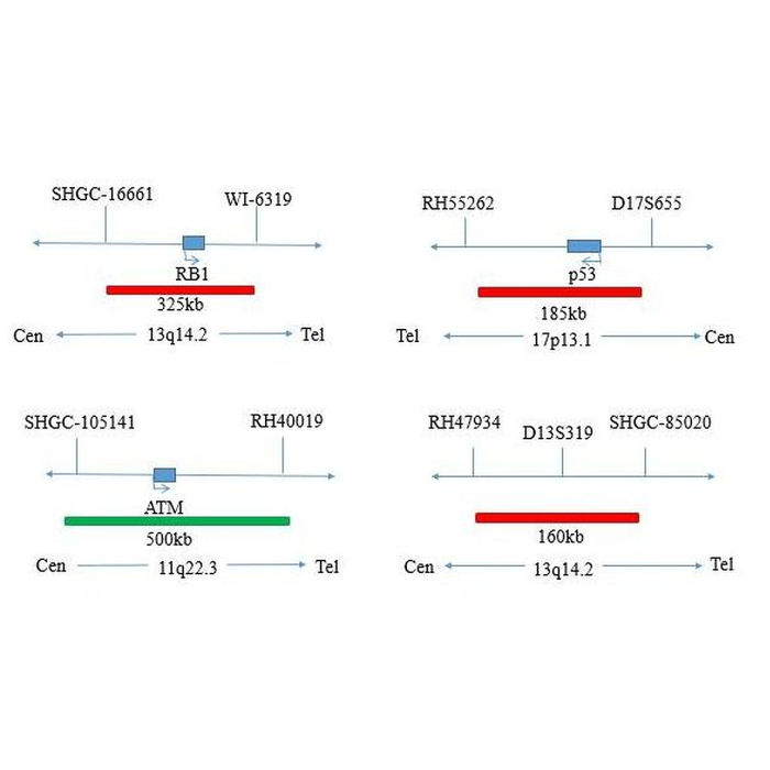
The kit consists of P53/CEP17 dual-color probe
|
Component name |
Specifications |
Quantity |
Main components |
|
P53/CEP17 dual-color probe |
100μL/Tube |
1 |
P53 Orange probe, CEP17 Green |
Intended use
This product is mainly used to detect chromosome and gene abnormalities in chronic lymphoblastic leukemia; The applicable population is patients suspected or diagnosed with chronic lymphocytic leukemia by routine clinical examination. Chronic lymphocytic leukemia (CLL) is a mature B-lymphocyte clonal proliferative tumor characterized by lymphocyte aggregation in peripheral blood, bone marrow, spleen and lymph nodes. Peripheral blood B lymphocytes with clinical diagnosis of persistence (3 months) ≥ 5×10⁹ /L (e.g. peripheral blood B cells <5×10⁹/L), Chronic lymphocytic leukemia is also diagnosed in patients with hematopenia caused by bone marrow infiltration or disease-related symptoms.
Fluorescence in situ hybridization showed that about 80% of patients with chronic lymphoblastic leukemia had chromosome abnormalities, and the most common deletion was in the long arm del of chromosome 13 (13q14.1); Chromosome 12 deletion or trisomy; Chromosome 17 short arm deletion del (17p). These abnormalities are of great significance for the diagnosis, differential diagnosis, treatment and prognosis of chronic lymphocytic leukemia. This kit is not clinically verified in combination with gene targeted therapeutic drugs, but only for gene detection performance. This kit is only applicable to the detection of chronic lymphocytic leukemia and provides doctors with auxiliary information for diagnosis.
Detection principle
Based on the fluorescence in situ hybridization technology, a nucleotide of the nucleic acid probe is labeled with fluorescein. The detected target gene is homologous and complementary with the used nucleic acid probe. After denaturation, annealing and renaturation, they can form a hybrid between the target gene and the nucleic acid probe. The detected gene is analyzed qualitatively, quantitatively or relatively under the microscope by the fluorescence detection system. The kit adopts orange probe labeled with orange fluorescein and green probe labeled with green fluorescein. The two probes can be combined with the target detection site by in situ hybridization.
Under normal conditions (without gene deletion and chromosome abnormality), it is displayed as two orange signals and two green signals under fluorescence microscope. When there is gene deletion, there will be a lack of green or orange signal. When there is chromosome polysomy, the probe signal will increase. Gene deletion and chromosome abnormalities were detected by this method, so as to provide reference basis for clinical identification, prognosis judgment and medication of leukemia patients.