HER2 Gene Amplification Probe Detection Kit (HER2)
HER2 Gene Amplification Probe Detection Kit - 100µL/10 Tests
ERBB2, commonly referred to as HER2, is amplified and/or overexpressed in 20-30% of invasive breast carcinomas. HER2-positive breast cancer is treated in a separate manner from other subtypes of breast cancer and commonly presents as more aggressive disease. Metastatic HER2-positive breast cancer is now commonly treated with HER2-targeted therapy. Apart from being amplified/overexpressed, ERBB2 activating mutations have been shown to have clinical importance in HER2-negative breast cancer. These mutations have shown sensitivity to the tyrosine kinase inhibitor neratinib, and highlight the importance of clinical sequencing efforts in treating breast cancer.
This gene encodes a member of the epidermal growth factor (EGF) receptor family of receptor tyrosine kinases. This protein has no ligand binding domain of its own and therefore cannot bind growth factors. However, it does bind tightly to other ligand-bound EGF receptor family members to form a heterodimer, stabilizing ligand binding and enhancing kinase-mediated activation of downstream signaling pathways, such as those involving mitogen-activated protein kinase and phosphatidylinositol-3 kinase. Allelic variations at amino acid positions 654 and 655 of isoform a (positions 624 and 625 of isoform b) have been reported, with the most common allele, Ile654/Ile655, shown here. Amplification and/or overexpression of this gene has been reported in numerous cancers, including breast and ovarian tumors. Alternative splicing results in several additional transcript variants, some encoding different isoforms and others that have not been fully characterized.
Intended Use
This kit uses fluorescence in situ hybridization to detect the HER2 gene amplification status in-vitro. The test sample is a paraffin embedded specimen of breast cancer tissue. The product has not been clinically validated in conjunction with HER2 targeted therapies, and its clinical testing capabilities have been confirmed by comparative trial studies with companion diagnostics that have targeted drug validation. The test results of the product should not be used as the sole basis for individualized treatment.
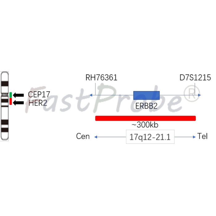
The clinician should make comprehensive judgment on the test results in combination with the patient's condition, drug indications, treatment response, and other laboratory testing indicators. Human epidermal growth factor receptor 2 (HER2, also known as Neu, ErbB-2, CD340, or p185) is a proto-oncogene HER2/neu located at the 17q12 of the human chromosome 17 long arm. Encoded, it is one of epidermal growth factor receptor (EGFR/ErbB) family members, has tyrosine kinase activity and is involved in signal transduction of cell growth and differentiation. The carcinogenic mechanism of HER2 oncogene includes inhibition of cell apoptosis, promotion of cell proliferation, increase of tumor cell invasiveness, and promotion of tumor angiogenesis and lymphangiogenesis. Breast cancer is one of the most serious malignant tumors that threaten women's health. Its incidence has been increasing year by year. HER2 gene amplification is found in 20% to 30% of diagnosed breast cancer patients. Patients with HER2-positive breast cancer often show high levels of tumor malignancy, poor treatment outcomes, and poor prognosis, but can benefit from specific HER2-targeted drug therapies. Therefore, the detection of HER2 gene amplification status in breast cancer patients is the precondition for the screening of HER2-targeted breast cancer patients and the curative effect prediction.
Product Main Components
The kit consists of FUS dual color probe
|
Component name |
Specifications |
Quantity |
Main components |
|
HER2/CEP17 dual color probe |
100μL/Tube |
1 |
HER2 Orange probe CEP17 Green probe |
Product Content
The kit is based on fluorescence in situ hybridization technology. A nucleic acid probe is labeled with fluorescein. The target gene is detected with homologous complementary to the nucleic acid probe used. Both after denaturation, annealing and renaturation, the hybrid of the target gene and the nucleic acid probe can be formed, and the qualitative, quantitative or relative positioning analysis of the gene to be measured under the microscope can be performed by the fluorescence detection system.
This kit uses the rhodamine fluorescein (RHO)-labeled orange probe (HER2 probe) to detect the HER2 gene, and the fluorescein isothiocyanate (FITC)-labeled green probe (CEP17 probe) to detect chromosome 17 centromere sequence can be used to bind two probes to the target detection area by in situ hybridization. The number of signals corresponding to the CEP17 probe reflects the number of chromosomes at the target area, and the number of signals from the HER2 probe reflects the copy number of the HER2 gene at the target site. By the ratio of the number of HER2 probes and the number of CEP17 probe signals, the amplified state of the HER2 gene in the tissue to be detected can be determined.