TERC gene amplification detection probe (TERC)
ERC Gene Amplification Probe Detection Kit - 100µL/10 Tests
Telomerase is a ribonucleoprotein polymerase that maintains telomere ends by addition of the telomere repeat TTAGGG. The enzyme consists of a protein component with reverse transcriptase activity, and an RNA component, encoded by this gene, that serves as a template for the telomere repeat. Telomerase expression plays a role in cellular senescence, as it is normally repressed in postnatal somatic cells resulting in progressive shortening of telomeres.
Deregulation of telomerase expression in somatic cells may be involved in oncogenesis. Studies in mouse suggest that telomerase also participates in chromosomal repair, since de novo synthesis of telomere repeats may occur at double-stranded breaks. Mutations in this gene cause autosomal dominant dyskeratosis congenita, and may also be associated with some cases of aplastic anemia.
Product Main Components
The kit consists of TERC/ CEP3 dual-color probes
|
Component name |
Specifications |
Quantity |
Main components |
|
TERC/ CEP3 dual color probe |
100μL/Tube |
1 |
TERC Orange probe, CEP3 Green probe |
Intended use
This kit uses fluorescence in situ hybridization to detect TERC gene status in vitro, and the test sample is cervical exfoliated cells. Cervical cancer is one of the most common malignant tumors in the female reproductive system. The incidence rate of cervical cancer is the second place in the world, which seriously threatens the health of women. Understanding the molecular basis of tumorigenesis and determining genetic markers by studying the genetic changes of cervical cancer is of great significance to early predict the prognosis of high-risk patients.
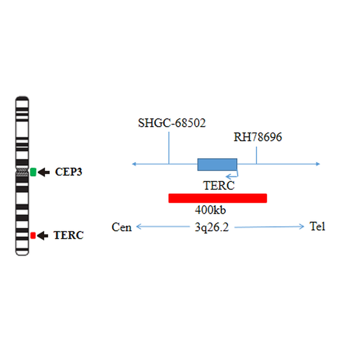
Studies have shown that the occurrence and development of cervical cancer is often accompanied by chromosome 3q amplification, located at 3q26 Human telomerase gene (hTERC) in region 3 can be amplified by chromosome 3q amplification. Studies have confirmed that the abnormal change of TERC is the necessary condition for the development of cervical intraepithelial neoplasia to cervical cancer, and the positive rate of TERC gene will increase with the increase of cervical lesion level.
Therefore, it has a certain predictive significance for the progression of cervical intraepithelial neoplasia. TERC gene detection can detect TERC gene amplification in cervical precancerous lesions, so TERC gene detection can be used for early diagnosis of cervical cancer, and TERC gene amplification level is positively correlated with tumor malignancy.
It is feasible to assist in the diagnosis of high-grade cervical intraepithelial lesions, and also has important clinical significance for the identification of surgical treatment indications, It can predict the development trend of cervical precancerous lesions to a certain extent, and can be a good supplement to the examination of cervical cell morphology and molecular cytogenetics, so as to provide a new way for the screening and prognosis evaluation of cervical cancer and precancerous lesions. This kit only verifies the detection performance of TERC gene. This kit is only applicable to the detection of TERC gene status and provides doctors with auxiliary information for diagnosis.
Detection principle
Fluorescence in situ hybridization is a technique for directly observing specific nucleic acids in cells in vitro. According to the principle of base complementary pairing, the specific DNA sequence is complementary to the target sequence in the cell. Due to the fluorescence of the probe, the hybrid probe and target DNA can be clearly observed under the fluorescence microscope under the appropriate excitation light. Hybridization includes the following steps: first, preprocess the samples according to the instructions; Secondly, DNA was denatured and hybridized with the probe; After hybridization, the excess unbound probes were washed away through a series of washing, and the nuclei were counterstained blue with DAPI (4,6-diamidino-2-phenylindole hydrochloric acid); Finally, the fluorescence signals from DAPI and the probe were observed under a suitable filter by fluorescence microscope.
The kit uses orange fluorescein labeled TERC probe to detect TERC gene, and green fluorescein labeled cep3 probe to detect the centromere sequence of chromosome 3. The two probes can be combined with the target detection site by in situ hybridization. The signal number corresponding to cep3 probe reflects the number of chromosome 3 at the target site, and the signal number of TERC probe corresponds to the status of TERC gene at the target site. The amplification status of TERC gene in the cells to be detected can be determined by the ratio of orange signal to green signal fluorescence number.