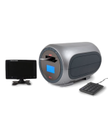
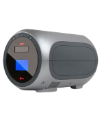
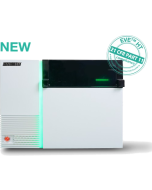
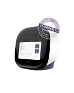
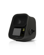
JuLI™ Stage Real-Time Cell History Recorder


JuLI™ Stage Real-Time Cell History Recorder
Cell biology is moving to video generation. Image, Video and Editing simultaneously and automatically.
JuLI™ Stage is designed to support cell biology researchers to approach kinetic images and data from start to end, saving time and allowing focus on more advanced and valuable work. Equipped with a fully automated x-y-z stage and multi-channel fluorescent colors, it acquires cell images and videos from various cell culture plates (up to 384-well) and dishes inside an incubator. It enables quantified cell confluence results with low variation as well as growth curve analysis using image-based analysis for bright field applications.
Key Applications
- Wound healing assays
- Apoptosis & Cytotoxicity monitoring
- Cell proliferation studies
- Fluorescence expression analysis
- 3D-spheroid culture monitoring
- Stem cell monitoring
- Cell growth monitoring
- Neuronal differentiation tracking
- Angiogenesis studies
- And more...
Main Functions
- Image capturing with high resolution
- Time-lapse recording capabilities
- Automated video making
- Image stitching for large samples
- Image & video editing tools included
- Multi-channel fluorescent colors (GFP, RFP, DAPI)
- Multi-well monitoring (up to 384 wells)
- Multi-position monitoring within plates
Key Features
- Incubator-compatible design (fits inside standard incubators)
- Fully automated X-Y-Z stage for precise positioning
- Interchangeable objective lenses (4x, 10x, 20x)
- Manual & auto focusing capabilities
- Compatible with various plates & vessels (dishes, flasks, multi-well plates)
- Integrated data management with all-in-one PC
- Real-time monitoring without removing samples from incubator
Application Videos
Browse time-lapse videos and images taken with the JuLI™ Stage to see how researchers around the world use this system for live cell imaging:
▶ JuLI Stage Overview Video
▶ Cell Migration Assay
▶ 3D Spheroid Monitoring
▶ Wound Healing Assay
▶ Fluorescence Imaging
▶ Cell Proliferation Analysis
Real-time, high-quality images and time-lapse videos are essential for modern cell biology research. JuLI™ Stage delivers automated imaging solutions for continuous monitoring of cell cultures.
| Light Source | Blue, Green, UV LED (Intensity adjustable) |
|---|---|
| Objective Lens | 4X, 10 X, 20 X + Digital ZoomSpec |
| Fluorescence | 3 fluorescence DAPI: Excitation 390/40, Emission 452/45 GFP: Excitation 466/40, Emission 525/50 RFP: Excitation 525/50, Emission 580LP |
| Camera | High-sensitivity monochrome CCD (Sony sensor 2/3”) 1,936 ⅹ1,456 pixels (2.8 M), 53 FPS, 14 bit |
| Stage | Automated X-Y-Z stage Inter-changeable vessel holder |
| Exported formats | Image: JPEG, TIFF, BMP, PNG Video: AVI Raw data: CSV |
| Image resolution | 2 M Pixels |
| PC | All-in-One touch screen desktop CPU: Intel Core i5-4590S Processor (Qual Core, 6MB,3.00GHz) OS: Genuine Windows 8.1 64bit (ENG) RAM: 8 GB (2x4 GB) 1600MHz DDR3L Memory HDD: 1TB 2.5” SATA (5,400 Rpm) 23” Full HD (1920 X 1080) with touch screen |
| Power Supply | 100 – 240 V - 50/60 Hz - 120 W |
| Dimensions | 429 (W) X 310 (D) X 324 (H) mm |
| Catalog Number | Product Name | Price | Qty |
|---|---|---|---|
| JSCT100 | JuLI Stage Scratcher (Scratcher Device, 1x Plate Rack, 2x Wash Rack) |
|
|
| JSCT100-2 | JuLI Stage Scratch Software |
|
|
| JSPT100 | JuLI Stage Spheroid Software |
|
|
| JS0100 | JuLI Stage Desktop PC |
|
|
| JMO100 | JuLI Stage PC Monitor / 24" Full HD |
|
|
| JP0150 | External Hard Disc Drive - 8TB(4TB x 2ea) |
|
|
| JO0004 | Objective Lens - 4X |
|
|
| JO0010 | Objective Lens - 10X |
|
|
| JO0020 | Objective Lens - 20X |
|
|
| JVH001 | Vessel Holder - Micro Slide (25 x 75mm) |
Out of stock
|
|
| JVH002 | Vessel Holder - Petri Dish (35mm) |
|
|
| JVH003 | Vessel Holder - Petri Dish (60mm) |
|
|
| JVH004 | Vessel Holder - Petri Dish (100mm) |
|
|
| JVH005 | Vessel Holder - T-Flask (25 & 75cm2) |
|